
Page 35 - 中国药房2021年11期
P. 35
红色 绿色 合并 表 2 各组 H9c2 心肌细胞中 ATP 酶活性测定结果(x±±
s,n=3)
Tab 2 ATPase activity in H9c2 cardiomyocytes of
正常对照组 each group(x±±s,n=3)
+
组别 Na -K -ATP酶,U/mg Ca -Mg -ATP酶,U/mg
+
2+
2+
正常对照组 38.45±2.50 26.77±2.08
模型组 17.80±2.21 ** 12.75±2.35 **
荭草花提取物低浓度组 22.37±3.14 14.41±4.01
荭草花提取物中浓度组 24.14±4.05 # 19.29±2.00 #
荭草花提取物高浓度组 26.27±1.98 ## 21.85±4.61 #
**
##
#
模型组 0.01 注:与正常对照组比较, P<0.01;与模型组比较,P<0.05,P<
* *
#
Note:vs. normal control group, P<0.01;vs. model group,P<
0.05,P<0.01
##
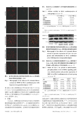
线粒体中Cyto c 14 kDa
荭草花提取物低浓度组 细胞质中Cyto c 正常对照组 模型组 荭草花提取 荭草花提取 荭草花提取 14 kDa
β-actin
42 kDa
物高浓度组
物低浓度组
物中浓度组
图 5 荭草花提取物对缺氧复氧损伤 H9c2 心肌细胞的
细胞质和线粒体中Cyto c蛋白表达影响的电泳图
荭草花提取物中浓度组 extract on protein expression of Cyto c in cyto-
Micrograph of the effects of P. orientale flower
Fig 5
plasm and mitochondria in H9c2 cardiomyo-
cytes with hypoxia-reoxygenation injury
表 3 各组 H9c2 心肌细胞的细胞质中 Cyto c/线粒体中
Cyto c比值、RISK信号通路相关蛋白磷酸化水平
荭草花提取物高浓度组 Tab 3 Ratio of Cyto c in cytosol to mitochondria,
及HIF-1α蛋白表达水平测定结果(x±±s,n=3)
phosphorylation level of RISK signaling path-
way related protein and protein expression of
HIF-1α in H9c2 cardiomyocytes of each group
图 4 荭草花提取物对缺氧复氧损伤 H9c2 心肌细胞 (x±±s,n=3)
MMP影响的显微图(×200) 细胞质中Cyto c/ p-ERK1/2/ HIF-1α/
组别 p-Akt/Akt
Fig 4 Micrograph of the effects of P. orientale flower 线粒体中Cyto c ERK1/2 β-actin
正常对照组 0.73±0.06 0.76±0.09 0.98±0.06 0.16±0.05
extract on MMP in H9c2 cardiomyocytes with
模型组 1.85±0.08 ** 0.20±0.07 ** 0.66±0.06 ** 0.92±0.04 **
hypoxia-reoxygenation injury(×200) 荭草花提取物低浓度组 1.57±0.06 # 0.32±0.03 # 0.91±0.06 ## 0.69±0.07 ##
荭草花提取物中浓度组 1.23±0.07 ## 0.52±0.05 ## 1.10±0.06 ## 0.48±0.08 ##
和 ERK1/2 蛋白的磷酸化水平均显著升高(P<0.05 或 荭草花提取物高浓度组 0.92±0.08 ## 0.70±0.05 ## 1.21±0.06 ## 0.35±0.04 ##
P<0.01);与荭草花提取物组比较,LY+荭草花提取物组 注:与正常对照组比较, P<0.01;与模型组比较,P<0.05,P<
#
**
##
细胞中Akt蛋白磷酸化水平和PD+荭草花提取物组细胞 0.01
中 ERK1/2 蛋白磷酸化水平均显著降低(P<0.05 或 P< Note:vs. normal control group, P<0.01;vs. model group,P<
* *
#
0.01),详见图7、表4。 0.05,P<0.01
##
4 讨论 损伤过程中,细胞内的氧化应激和钙离子诱导的线粒体
CoCl2是一种模拟缺氧条件的化学试剂,常被用来 功能障碍驱动了细胞的损伤和凋亡 [21-23] ,其中ROS是氧
[21]
制备细胞缺血缺氧损伤模型 。在本课题组前期研究 化应激中破坏机体氧化-抗氧化系统平衡的主要根
的基础上 ,本研究采用 800 μmol/L CoCl2复制 H9c2 心 源 。本研究发现,缺血缺氧损伤H9c2心肌细胞的细胞
[24]
[3]
肌细胞缺氧复氧损伤模型。在体内外的细胞缺氧复氧 核固缩、致密浓染,可见大量凋亡小体;细胞中ROS水平
中国药房 2021年第32卷第11期 China Pharmacy 2021 Vol. 32 No. 11 ·1309 ·