
Page 43 - 《中国药房》2021年第1期
P. 43
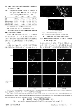
表1 OGD后复氧不同时间的各组细胞中LDH含量比
             较(x±±s,n=8,U/L)
        Tab 1 Comparison of LDH content in astrocyte in
               each group after different time of reoxyge-
               nation following OGD(x±±s,n=8,U/L)
        组别             复氧0 h    复氧3 h    复氧6 h    复氧12 h
        对照组           29.42±3.78  32.01±7.14  37.87±7.74  36.43±10.5  A.对照组                 B.模型组
        模型组           110.99±17.06 *  161.51±30.25 *  344.65±34.94 *  436.64±55.29 *
        小续命汤含药血清低浓度组  90.22±15.23  122.61±29.22  190.67±21.79 #  253.55±32.23 #
        小续命汤含药血清中浓度组  78.90±19.43  89.06±17.53  141.1±18.89 #  218.56±30.32 #
        小续命汤含药血清高浓度组  65.96±17.90  86.05±18.42  121.23±26.47 #  177.26±27.30 #
                                         #
                        *
           注:与对照组比较,P<0.05;与模型组比较,P<0.05
                                              #
           Note:vs. control group,P<0.05;vs. model group,P<0.05
                           *
        3.3  小续命汤含药血清对OGD损伤模型大鼠星形胶质                                       C.小续命汤含药血清高浓度组
        细胞中MnSOD水平的影响                                          注:箭头处表示DCFH-DA荧光标记的ROS
            对照组细胞中平均光密度为 46.28±11.57;模型组                       Note:the arrow indicates the ROS labeled with DCFH-DA
        中 平 均 光 密 度 为 4.63 ± 1.72,显 著 低 于 对 照 组(P<              图1 各组细胞中ROS的荧光显微图(×400)
        0.05);小续命汤含药血清高浓度组细胞中平均光密度为                        Fig 1 Fluorescence micrograph of ROS in astrocyte
        32.79±15.7,显著高于模型组(P<0.05),详见图2。                          of each group(×400)
        4 讨论                                               参、桂枝、黄芩、川芎、芍药各一两,防风一两半,生姜五
            小续命汤治疗缺血性卒中的历史可追溯至我国南                          两,附子大者一枚”,因治疗卒中效果显著,被当时众医
                                                                            [18]
        北朝时期陈延之的《小品方》,曰“甘草、麻黄、防己、人                         奉为“诸汤之最要” 。后被我国唐代孙思邈收录于《备


                对照组


                模型组


         小续命汤含药血清高
         剂量组


                             DAPI                 GFAP                  MnSOD               Merge重叠
           注:第1列为蓝色荧光标记的DAPI,表示细胞核;第2列为红色荧光标记的GFAP,表示星形胶质细胞;第3列为绿色荧光标记的MnSOD;第4
        列为前3列的重叠图,呈黄色荧光,表示MnSOD在星形胶质细胞内的表达定位
           Note:the first column is DAPI labeled with blue fluorescence,indicating the nucleus;the second column is GFAP labeled with red fluorescence,
        indicating astrocytes;the third column is MnSOD labeled with green fluorescence;the fourth column is the overlapping diagram of the first three co-
        lumns,showing yellow fluorescence,indicating the expression and localization of MnSOD in astrocytes
                                   图2   各组细胞中MnSOD的免疫荧光显微图(×400)
                     Fig 2 Immunofluorescence micrograph of MnSOD in astrocyte of each group(×400)


        中国药房    2021年第32卷第1期                                                China Pharmacy 2021 Vol. 32 No. 1  ·37 ·